Localization
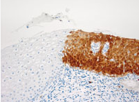
Positive CINtec p16 Histology staining is characterized as diffuse, continuous staining of cells of the basal and parabasal cell layers of the squamous cervical epithelium, with or without staining of cells of intermediate to superficial cell layers. This staining pattern is representative of overexpression of the p16INK4a protein within the cervical epithelium (Panel A).
In some specimens, nuclear expression may be faint or undetectable, but nuclear staining is not required to interpret the p16INK4a staining. Therefore, both nuclear and/or cytoplasmic immunostaining should be taken into account when assessing the p16INK4a staining pattern.
Negative staining is demonstrated by either focal staining or no staining of the cervical epithelium.
A focal CINtec p16 Histology staining pattern is demonstrated by non-continuous staining of isolated cells or small cell clusters, particularly not of the basal and parabasal cells of the cervical squamous epithelium. The focal staining pattern is interpreted as a negative CINtec p16 Histology result (Panel B).
In addition, no p16INK4a IHC staining of cells within the cervical squamous epithelium results in a negative CINtec p16 Histology result (Panel C).
Description
CINtec p16 Histology is an immunohistochemistry assay for the qualitative detection of the p16INK4a protein on formalin-fixed, paraffin-embedded tissue sections prepared from cervical biopsies. It is intended for use in medical pathology laboratories to provide adjunctive information after initial diagnosis has been made by established diagnostic methods.
This product should be interpreted by a qualified pathologist in conjunction with histological examination, relevant clinical information and proper controls.
The product is intended for in-vitro diagnostic (IVD) use.


CINtec p16 Histology staining of cervical biopsy specimens with the OptiView Detection Kit on the VENTANA BenchMark ULTRA IHC / ISH automated staining instrument. Panel A: Positive CINtec p16 Histology staining in a diffuse, continuous staining pattern comprising the basal and parabasal cell layers of the squamous cervical epithelium. Panels B and C: Negative staining patterns represented by focal staining (B) and no staining of the squamous cervical epithelium (C).